Mitral Valve Scallops
- cardiacmrihub
- Nov 24, 2025
- 3 min read
Updated: Dec 1, 2025
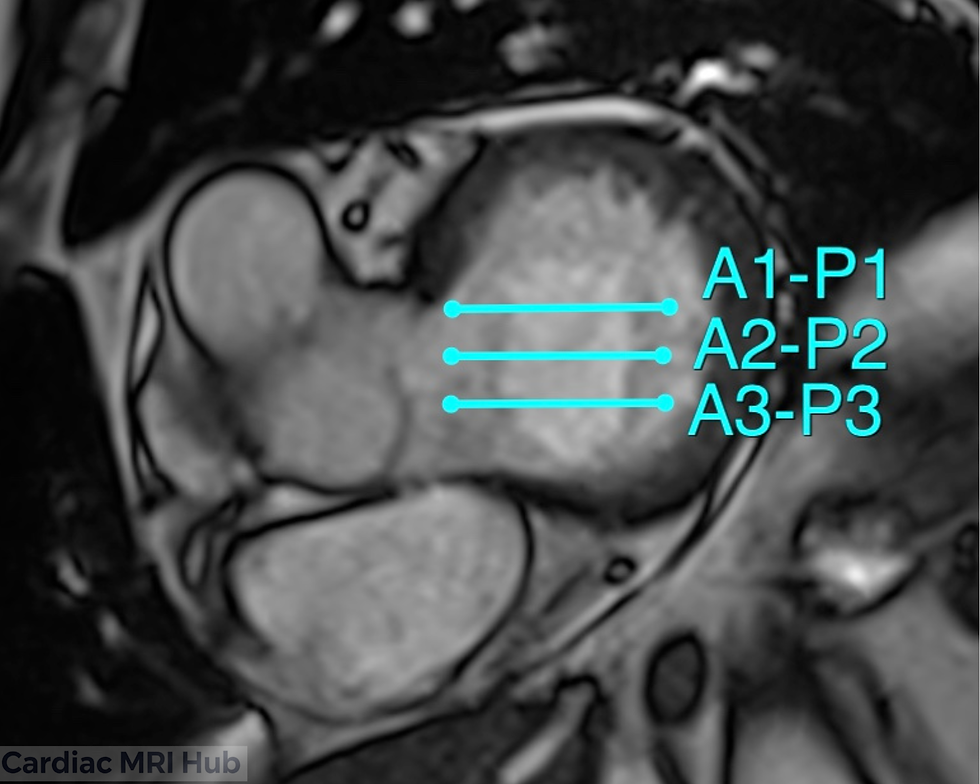
The mitral valve scallops view is an essential imaging technique in cardiac MRI that provides detailed visualization of the mitral valve's complex anatomy. Using the Carpentier classification system, the mitral valve is divided into six distinct segments or scallops, three anterior (A1, A2, A3) and three posterior (P1, P2, P3) which allows for precise localization of valvular pathology. This imaging approach is particularly valuable for evaluating mitral valve prolapse, regurgitation, and structural abnormalities, making it crucial for pre-surgical planning and guiding interventional procedures. By systematically imaging each scallop segment, technologists can provide clinicians with comprehensive information about leaflet motion, coaptation defects, and chordal apparatus integrity.
Mitral Valve Scallops Planning


Planning for Mitral Valve Scallops View in Cardiac MRI
Key Considerations for Planning
Orientation: The scallops view is typically acquired from a plane that optimally captures the mitral valve's scallops, ensuring clear visualization of the valve leaflets and their relationship to the surrounding structures.
Slice Thickness: A thinner slice thickness is recommended to enhance the resolution and detail of the mitral valve structures.
Field of View: The field of view should encompass the entire mitral valve apparatus, including the annulus, leaflets, and chordae tendineae, to avoid missing critical features.
Timing: Image acquisition should be timed with the cardiac cycle, ideally during diastole, to optimize visualization of the mitral valve's opening and closing dynamics.
Contrast Agents: The use of contrast agents may be beneficial to improve the delineation of the mitral valve structures and enhance diagnostic accuracy.
Clinical Applications
Assessment of mitral valve morphology and function.
Identification of mitral valve prolapse or regurgitation.
Evaluation of associated left atrial and ventricular changes.
Monitoring of mitral valve disease progression over time.
Key Features of the Mitral Valve Scallops View
Mitral Annulus: Evaluation of annular dimensions, shape, and calcification to assess for annular dilatation or restriction, which can contribute to mitral regurgitation.
Mitral Leaflets: Detailed analysis of the anterior and posterior leaflets, including their motion and any structural abnormalities.
Scallop Segmentation: Systematic evaluation of each individual scallop (A1, A2, A3, P1, P2, P3) to precisely localize areas of prolapse, restricted motion, or perforation using the Carpentier classification.
Coaptation Zone: Evaluation of the leaflet coaptation line and length to detect coaptation defects, malcoaptation, or excessive coaptation depth that may indicate prolapse or restriction patterns.
Papillary Muscles: Assessment of papillary muscle positioning, alignment, and morphology to identify dysfunction or displacement that may affect leaflet coaptation.
Chordae Tendineae: Assessment of the chordae tendineae for rupture or elongation, which may impact valve function.
Conclusion
The mitral valve scallops view is a fundamental component of comprehensive cardiac MRI protocols, enabling precise anatomical assessment and functional evaluation of the mitral valve apparatus. By systematically imaging each scallop segment using the Carpentier classification, MRI technologists provide essential diagnostic information that guides clinical decision-making for patients with mitral valve disease. Proper planning techniques, including optimal slice orientation, appropriate timing within the cardiac cycle, and adequate field of view, ensure high-quality images that accurately depict leaflet morphology, coaptation dynamics, and associated structural abnormalities. Mastery of this imaging approach enhances the value of cardiac MRI in pre-procedural planning, surgical guidance, and longitudinal monitoring of mitral valve pathology.


Comments